By Bhavana Kunkalikar May 22 2023 Reviewed by Benedette Cuffari, M.Sc.
In a recent study published in the journal Nature Communications, researchers explore the relationship between gout and neurodegenerative disease susceptibility.

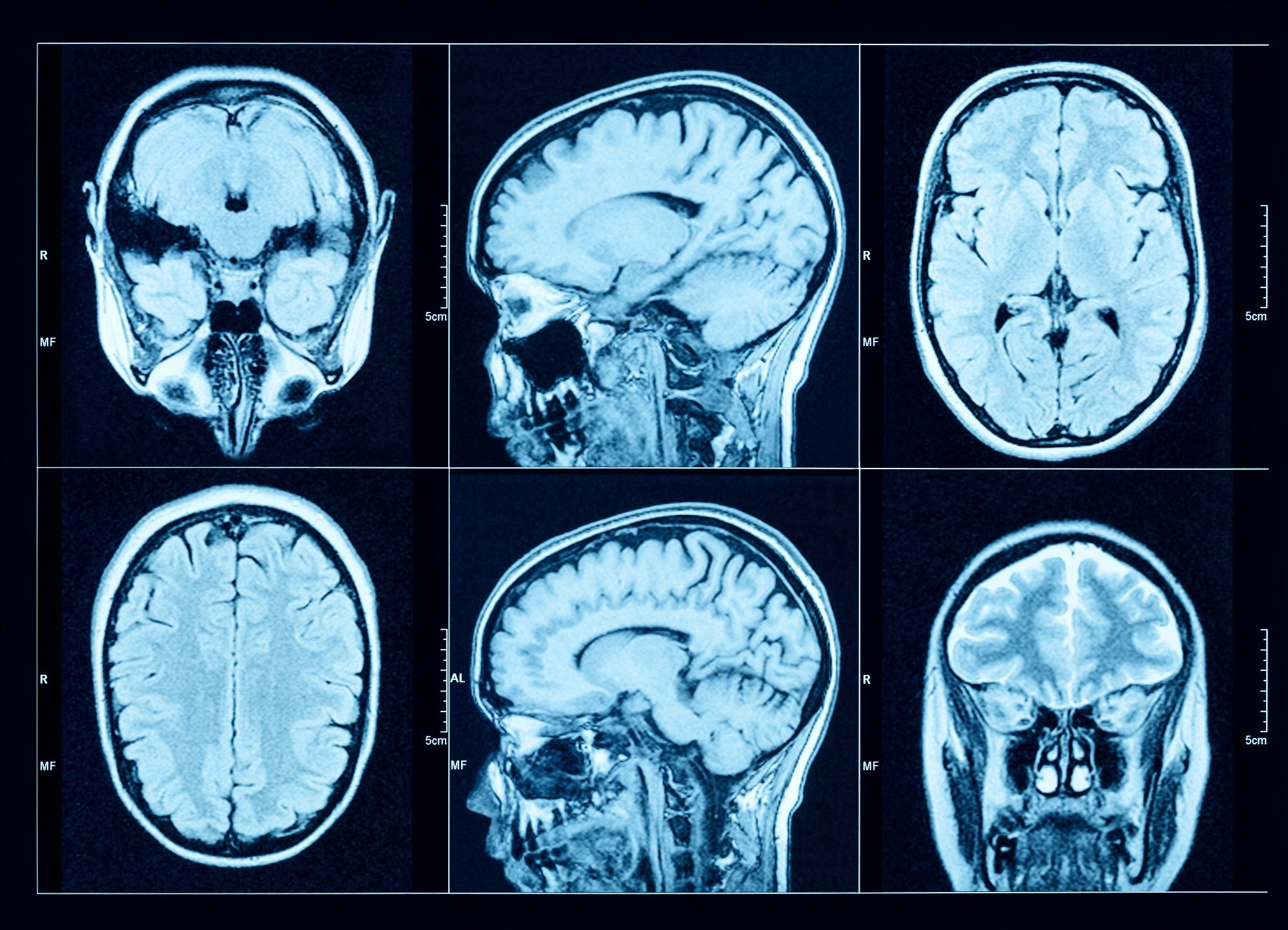
Study: Association of gout with brain reserve and vulnerability to neurodegenerative disease. Image Credit: Triff / Shutterstock.com
What is gout?
Gout, which is often referred to as hyperuricemia, is the most prevalent form of inflammatory arthritis, as it affects between 1% and 4% of the population. Gout is the result of monosodium urate crystal deposition in the joints and peri-articular tissues, which subsequently leads to an inflammatory reaction, swelling, and pain.
Recent studies have made conflicting observations regarding the relationship between gout and neurodegenerative diseases. For example, some observational studies have reported that gout is associated with a reduced risk of dementia, especially Alzheimer’s disease, whereas Mendelian randomization studies have not confirmed these findings. A history of gout has also been shown to increase the risk of stroke.
These contradictory findings emphasize the need for more studies, particularly those involving brain structure analysis, to better understand the relationship between gout and neurodegenerative diseases.
About the study
Study participants were recruited from the United Kingdom Biobank (UKB) study, which enrolled 40 to 69-year-old volunteers between 2006 and 2010. A subset of these patients underwent imaging, including magnetic resonance imaging (MRI) of the brain.
Diagnostic criteria for gout were derived algorithmically from UKB baseline assessment information collection. All participants’ serum urate levels were estimated at the beginning of the study.
The team utilized 2,138 summary image-derived phenotypes (IDPs) that represented different estimates of brain structure using T1-weighted and T2-weighted-FLAIR structural imaging, diffusion MRI, and susceptibility-weighted MRI. FMRIB software library voxel-based morphometry (FSL-VBM) was used to determine the precise spatial distribution of relationships between gout and gray matter volume throughout the brain.
MRI results were used to determine whether causal relationships could explain the observed associations with brain structure. One-sample (gout) and two-sample (urate) linear MRI assessments utilizing summary statistics obtained from European participants were also performed.
Variant harmonization guaranteed that the correlation between single nucleotide polymorphisms (SNPs) and exposures, as well as SNPs and outcomes, described the same allele. Several robust MRI techniques were employed to evaluate the consistency of each causal inference.
Gout increases the risk of dementia and Parkinson’s
A total of 11,735 participants with gout were included in the current study, 1,165 of whom underwent brain imaging. About 31% of all gout patients were currently being treated with urate-lowering therapy (ULT).
Most of the patients with gout were older and males. Notably, urate levels in male gout patients were positively correlated with alcohol intake and lower socioeconomic status.
During the follow-up period, 3,126 participants reported dementia, and 16,422 died, with gout patients twice as likely to die as compared to controls.
Urate levels were inversely associated with global brain, grey matter, white matter, and high cerebrospinal fluid volumes. In fact, gout was found to exert the same impact on global grey matter volume as that which is observed when comparing the brain scans of a healthy individual to those of an individual who is two years older.
Some of the specific gray matter regions of the brain that were affected by gout included the cerebellum, pons, and midbrain. Likewise, white matter regions, including the fornix, exhibited higher mean diffusivity and lower fractional anisotropy in gout patients.
A history of gout and high urate serum levels were also associated with a greater degree of iron deposition in the bilateral putamen and caudate, both of which are basal ganglia structures. High iron levels within these brain structures may be due to gout-related inflammatory processes or poor urinary iron excretion, as controlling for renal function was found to reduce this association.
Gout was positively associated with dementia, particularly vascular dementia, with the highest risk of a dementia diagnosis occurring in the first three years following a gout diagnosis. Additionally, gout increases the risk of both Parkinson’s disease and probable essential tremors.
Conclusions
The current study is the first of its kind to correlate neuroimaging studies with gout, with its findings providing evidence for a causal involvement of gout in dementia and other neurodegenerative disease. Nevertheless, the researchers did not observe any classical imaging markers of Alzheimer’s disease or vascular dementia in the brains of gout patients.
Taken together, the study findings are important for clinicians treating gout patients, as prophylactic treatments for gout may also reduce the risk of these patients developing neurodegenerative disorders in the future. These observations may also provide new insights into the different pathways involved in the pathogenesis of neurodegenerative diseases that can be used to identify novel therapeutic targets.

Leave a Reply