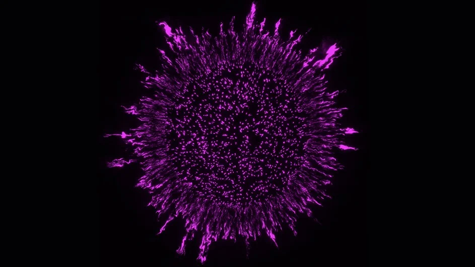
Bacteria are traditionally imagined as single-cell organisms, spread out sparsely over surfaces or suspended in liquids, but in many environments the true bacterial mode of growth is in sticky clusters called biofilms.
Biofilm formation can be useful to humans—it is integral, for example, to producing kombucha tea. But it is more often problematic because it makes it more difficult to control bacterial growth: When bacterial cells produce a biofilm, it acts as a shield against outside invaders, making the bacteria more tolerant to antibiotics.

Image credit: Columbia University
Until recently, researchers had assumed that bacteria were arranged somewhat randomly in biofilms, insofar as they had thought about the question of biofilm structure at all. But new research from Biology Professor Lars Dietrich’s lab shows that bacteria that form biofilms actually have a highly structured arrangement within those slimy matrices.
Their unexpected finding could pave the way for developing new drugs that better target antibiotic-resistant bacteria.
“There’s a yin-yang trade-off for bacteria that form biofilms, since the biofilm guards against antibiotics and other threats, but also prevents food from entering and feeding the system,” said Dietrich, a lead author on the paper. “This research gives us an important foundation for understanding how to affect bacterial-cell arrangement and assess how to make them more susceptible to antibiotics.”
The new study, published in the journal PLOS Biology, details research conducted in professor Dietrich’s lab, spearheaded by graduate student Hannah Dayton. The paper looked specifically at an important, common pathogen, called Pseudomonas aeruginosa.
The team used scanning electron microscopy and fluorescence microscopy paired with cell labeling to conduct their research. They found that P. aeruginosa cells in biofilms are packed lengthwise and arranged perpendicularly to their growth substrate, the material that the bacteria live on and that contains the substance they are eating to survive and grow. They also found that mutations that modify the bacterial cell surface disrupt this arrangement.
When they tested the effects of a sugar added from the outside to a fully formed biofilm, they observed that its distribution was affected by the biofilm anatomy. Mutant bacteria with a disordered cellular arrangement were more responsive to added sugar or antibiotic in specific areas within the biofilm, also known as subzones. Finally, they showed that changes in biofilm anatomy shift the location of peak metabolic activity within the structure.
Together, these observations indicate that biofilm microstructure is a property that can be tuned to influence the metabolism of resident bacterial subpopulations and affect the overall survival of the group. The findings have implications for our approaches to treating infections caused by P. aeruginosa and other biofilm-forming pathogens.
The research was conducted in collaboration with the research groups of Wei Min, a Columbia chemistry professor; Raju Tomer, a Columbia biology professor; Jasmine Nirody, professor at the University of Chicago; and Anuradha Janakiraman, professor at the City University of New York (CUNY).
“The arrangement of cells is generally an underappreciated aspect of biofilm formation,” said Dayton. “We now know that it allows bacteria in biofilms to control their physiological states and has consequences for their survival during antibiotic treatment.”
“This is a promising development for the pernicious and growing problem of antibiotic resistant bacteria,” Dietrich said.
Source: Columbia University

Leave a Reply