by Cedars-Sinai Medical Center

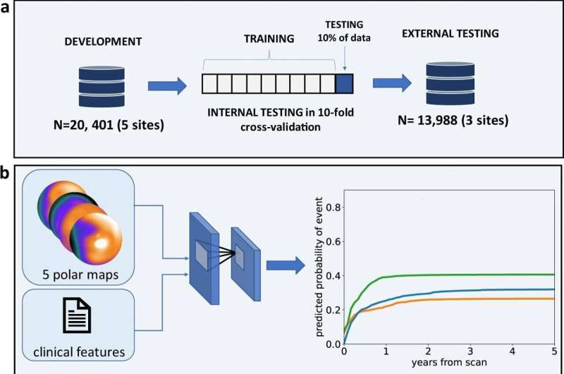
Deep learning enabled time-to-event outcome prediction after cardiac imaging – study overview. a A time-to-event deep learning model was trained (left) using data from the 5 sites of the REFINE SPECT registry (n = 20,401), then tested internally in a 10-fold cross-validation regimen (middle) and tested in 3 external sites (n = 13,988) (right); b The time-to-event model uses 5 SPECT polar maps and 15 clinical features as inputs (left) and predicts time-dependent probability of death (orange line), ACS (green line), and revascularization (blue line); c The performance of the model (left) is analyzed using cumulative dynamic area under the receiver-operating curves (cAUC). Red line represents the time-to-event model and blue line represents perfusion abnormality. The explanation of the prediction is visualized as a waterfall plot with blue arrows representing features that decrease the risk and red arrows representing the features that increase the risk (right); ACS acute coronary syndrome, AUC area under the receiver operating characteristics curve, TPD total perfusion deficit, PCI percutaneous coronary intervention, CI confidence intervals. Credit: npj Digital Medicine (2023). DOI: 10.1038/s41746-023-00806-x
What if your physician could predict if—or when—you might experience a heart attack, cardiac arrest or another heart-related problem?
Investigators are one step closer to achieving this breakthrough in preventive health care and offering patients personalized predictions of their heart health thanks to a novel deep-learning tool and artificial intelligence (AI) algorithm developed at Cedars-Sinai.
Investigators say their findings, published today in the journal npj Digital Medicine, might be an effective way to engage patients in their own health care.
“Using a specific type of AI trained to interpret images of the heart and developed at Cedars-Sinai, we could both predict the chance of cardiac events—like death, heart attack, or the need for urgent treatment of the heart vessels—and show how the likelihood of these adverse events changes over time,” said Piotr Slomka, Ph.D., director of Innovation in Imaging at Cedars-Sinai and a research scientist in the Division of Artificial Intelligence in Medicine and the Smidt Heart Institute.
To do this, the AI tool was trained to collect and interrogate basic clinical data like each patient’s age, gender, weight, heart rate, and blood pressure, as well as to interpret images of the heart that show blood flow to the heart muscle, and how the heart expands and contracts.
“This general patient data, together with heart imaging, is what the deep-learning platform uses to make cardiac health predictions,” said Slomka, senior author of the study.
The predictions are produced in a graph format that indicates individual risk for patient death, heart attack, or requirement for an invasive cardiovascular intervention—such as a stent or bypass surgery—over the period of several years. Slomka says the graphs are simple to understand and can be reviewed by both medical professionals and patients.
“Doctors and patients can use these graphs to track how risk changes over time and to identify individual risk factors,” said Slomka. “They can also interactively modify certain risk factors to see how it impacts a patient’s particular risk.”
Conceptually, this novel research could have broad implications, says Sumeet Chugh, MD, director of the Division of Artificial Intelligence in Medicine and the Pauline and Harold Price Chair in Cardiac Electrophysiology Research.
“AI algorithms of this nature could enable physicians to communicate more personalized information regarding potential timing of imminent heart disease events, allowing patients to engage more meaningfully in the shared decision-making process,” said Chugh, director of the Center for Cardiac Arrest Prevention in the Smidt Heart Institute. “Even more importantly, this tool has the potential to lend data-led, appropriate urgency to heart disease prevention efforts by both patients and providers.”
Slomka and his team of investigators plan to test these tools in clinical trials at Cedars-Sinai in the near future.
“Our ultimate goal is to offer such interactive tools online if images and clinical data are uploaded,” said Slomka.

Leave a Reply