by Thamarasee Jeewandara , Medical Xpress

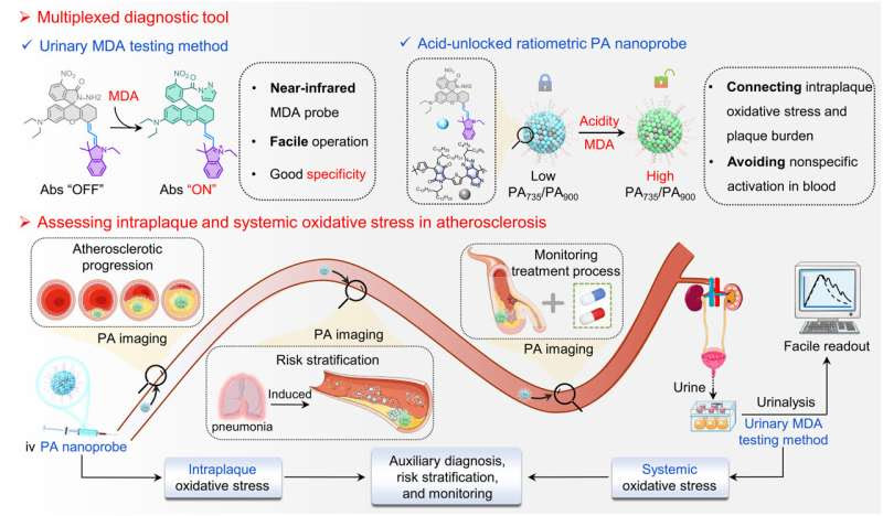
A multiplexed diagnostic tool for detecting intraplaque and urinary MDA. We design a multiplexed diagnostic tool that includes two functions (photoacoustic imaging and urinalysis), for assessing intraplaque and urinary MDA and reliably assessing intraplaque and systematic oxidative stress in atherosclerosis. Credit: Science Advances, DOI: 10.1126/sciadv.adh1037
Oxidative stress is fundamental to the development of diverse pathological conditions including atherosclerosis. However, the effect of oxidative stress on atherosclerosis remains to be known.
In a recent report in Science Advances, Yuan Ma and a team of scientists in chemistry, cardiology and medicine at the Hunan University, China, developed a multiplexed diagnostic instrument with photoacoustic imaging and urinalysis to determine intraplaque and urinary malondialdehyde (MDA) as well recognized end-products of oxidative stress.
The team conducted molecular design to develop the first, near-infrared malondialdehyde-responsive molecule (abbreviated MRM). Using a photoacoustic nanoprobe, the team reported intraplaque malondialdehyde as a measure reflecting the plaque burden. This MRM molecule detected urinary malondialdehyde with excellent specificity.
The outcomes showed a significant difference in urinary MDA between healthy adults and atherosclerotic patients. The multiplexed diagnostic instrument can detect both intraplaque and systemic oxidative stress levels during atherosclerotic plaque progression and drug treatment in atherosclerotic mice as a promising auxiliary diagnosis measure.
The pathology of atherosclerosis and measuring it via oxidative stress in the clinic
Cardiovascular and cerebrovascular diseases are a primary cause of morbidity and mortality worldwide, and atherosclerosis plays a significant role at all stages.
The formation of plaque is a hallmark of atherosclerosis in chronic progressive lesions, alongside an unpredictability of plaque rupture contributing to severe cardiovascular and cerebrovascular events. As a result, evaluating plaque characteristics is significant to reduce morbidity. Nevertheless, the accurate prediction of plaque stage and location remains challenging to determine the noninvasive and reliable methods underlying high-risk plaques.
Oxidative stress is a significant biomarker of atherosclerosis at present, induced by excess reactive oxygen species. Chronic and acute inflammation of vascular walls can also similarly increase oxidative stress to contribute towards the development of atherosclerosis.
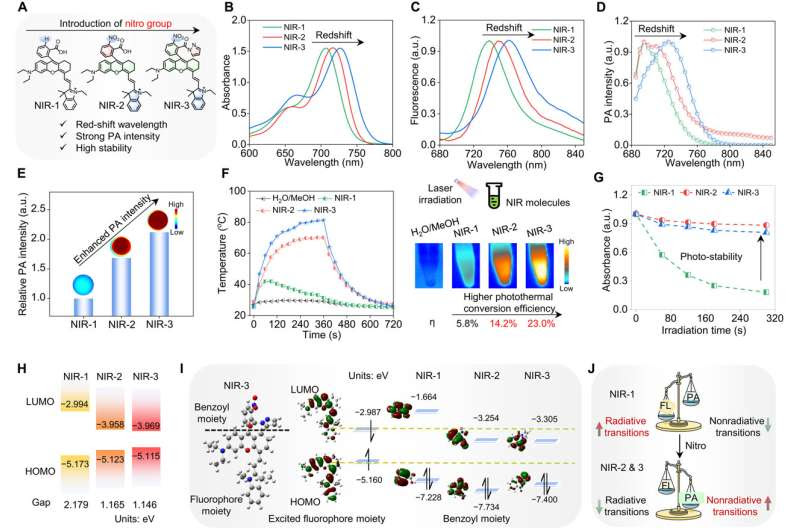
Oxidative stress biomarker triggered multiplexed instrument to diagnose atherosclerosisDesign, properties, and theoretical calculation of near-infrared molecule. (A) Chemical structures of NIR-1, NIR-2, and NIR-3. (B to D) Absorption (B), fluorescent (C), and PA (D) spectra of NIR-1, NIR-2, and NIR-3 in methanol (MeOH). (E) Relative PA intensity of NIR-1, NIR-2, and NIR-3 at 730 nm. (F) Heating (0 to 360 s) and cooling (360 to 720 s) operation, photothermal imaging, and photothermal conversion efficiencies of NIR-1, NIR-2, and NIR-3 in H2O/MeOH (v/v = 7:3) under 660-nm laser irradiation (power density, 1.6 W/cm2). (G) The photo-stability of NIR-1, NIR-2, and NIR-3 and normalized absorption of those molecules with 660-nm laser at a power density of 0.8 W/cm2 for 5 min. (H) HOMO/LUMO (highest occupied molecular orbital/lowest unoccupied molecular orbital) energy levels and energy gaps of NIR-1, NIR-2, and NIR-3. B3LYP exchange is functional using 6-31G basis sets using a suite of Gaussian 09. (I) Optimal structures of NIR-3, frontier orbital diagrams of the benzoyl moiety, and excited fluorophore moiety of NIR-1, NIR-2, and NIR-3. The orbital energies of those molecules were performed using the Gaussian 09 program at the B3LYP/6-31G level. (J) Balance of the radiative (fluorescence, FL) and nonradiative (photoacoustic, PA) transitions of NIR-1, NIR-2, and NIR-3. a.u., arbitrary units. Credit: Science Advances, doi: 10.1126/sciadv.adh1037
A photoacoustic nanoprobe from atherosclerotic mice to clinical use
Elevated levels of intraplaque oxidative stress can cause plaque vulnerability. As a result, the process of evaluating intraplaque oxidative stress can assist biologists and clinicians to identify plaque. Since traditional medical imaging methods are limiting to assess intraplaque and oxidative stress in atherosclerosis, Ma and colleagues sought to introduce a reliable instrument in clinic practice.
To accomplish this, they recognized malondialdehyde (MDA)—the product of lipid peroxidation as an ideal oxidative stress biomarker due to its sufficient stability. The team designed an MDA-responsive molecule (MRM) as a multiplexed diagnostic instrument.
The method conferred two functions—photoacoustic imaging and urinary malondialdehyde testing. The team used the photoacoustic nanoprobe in atherosclerotic mice to demonstrate its reliability in detecting oxidative stress and plaque characteristics in an atherosclerotic mouse model, with potential for auxiliary diagnostics and risk assay of atherosclerosis in clinical use.

Design, properties, and theoretical calculation of near-infrared molecule
Photoacoustic imaging agents are fundamental to improve the sensitivity of detection and to develop activatable probes. The team optimized the available photoacoustic agents for their medical imaging applications by introducing nitro groups to hemocyanin fluorescent molecules to improve the weak photoacoustic signal and photobleaching property of the fluorophore with near-infrared absorption and fluorescence emission.
Thereafter, the team measured the photothermal conversion capability of near infrared 1-to-3 in direct relation to photoacoustic intensity. To indicate improved photostability by introducing the nitro group, which inhibited the radiative transition process.
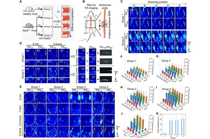
Oxidative stress biomarker triggered multiplexed instrument to diagnose atherosclerosisImaging the progression of atherosclerotic in living mice. (A) Scheme for construction of different progression of atherosclerosis and imaging procedure. Group 1: healthy mice; groups 2 to 4: ApoE−/− mice were fed with high-fat diet (HFD) for 10, 13, and 16 weeks. (B) Graphical views of PA imaging of living mice. Black area: scanning position; black line: scanning positions A to E for data analysis. (C) PA images of different scanning positions (A to E) in group 1 and group 4 mice after injection of PA nanoprobe. Aortic regions are depicted by an arrow. (D) PA images of groups 1 and 4 mice 5 min and 5 hours after injection of PA nanoprobe from transverse (XZ) and coronal (XY) plane. PA735/PA900 images from sagittal (YZ) plane of groups 1 and 4 mice 5 min and 5 hours after injection of the PA nanoprobe. The images of PA735/PA900 ratios were obtained via the PA735 images divided by PA900 images using Image-Pro Plus. (E) PA images of groups 1 to 4 mice after injection of PA nanoprobe. (F to I) Quantification of normalized PA735/PA900 ratios of group 1 (F), group 2 (G), group 3 (H), and group 4 (I) at different time points and scanning positions after injection of PA nanoprobe (three mice per group). (J) Quantification of normalized PA735/PA900 ratios of groups 1 to 4 at different time points after injection of PA nanoprobe (three mice per group, data for five scanning positions per mouse, means ± SD). (K) Relative total cholesterol content for groups 1 to 4 mice. Credit: Science Advances, doi: 10.1126/sciadv.adh1037
Design and response of malondialdehyde-responsive molecules
Since existing MDA probes exhibit short-wavelength fluorescence emission that leads to reduced tissue penetration, high background interference and a lower signal-to-noise ratio, Ma and the team used near-infrared-2 as the photoacoustic scaffold with near-infrared absorption and excellent photoacoustic properties. They also designed MDA-responsive molecule coupling hydrazine with benzoic acid to ultimately ‘turn-on’ absorption, fluorescence, and the photoacoustic signal.
The scientists showed how the introduction of the nitro group greatly improved the response of MDA-responsive molecules to malondialdehyde under acidic conditions. The outcomes highlighted the sensitivity of the malondialdehyde molecule to detect MDA in weak acidic conditions. The researchers optimized urinary MDA testing methods as one of the indicators of systemic oxidative stress to gain insights on atherosclerotic progression.
Testing urinary malondialdehyde levels
While urinary malondialdehyde (MDA) can be detected with the commercial kit where thiobarbituric acid reacts with the compound to form TBA-MDA, this can interfere with the accurate detection of urinary malondialdehyde. The team therefore developed a more specific testing method with malondialdehyde-responsive molecule to accurately determine the constituent of interest, without interference from external analytes.
Since malondialdehyde already exists in blood, the current probe can be activated in blood before it reaches the plaques to result in a false imaging signal. To prevent this, the team designed an acid-unlocked ratiometric photoacoustic nanoprobe. In its mechanism of action, the photoacoustic nanoprobe can be coactivated by acidity and the presence of malondialdehyde within the plaque, while the nanoprobe silently circulates blood due to the neutral pH of blood.
The scientists conducted reliable analysis of plaque characteristics ex vivo. Elevated levels of oxidative stress within the plaques can lead to local proteolysis, plaque rupture, and thrombosis formation in atherosclerotic arteries, to trigger cardiovascular events. It is important to study the generation of malondialdehyde as an endpoint since it is a biomarker of reactive oxygen-species driven elevated oxidative stress during atherosclerosis. The work confirmed the reliability of the photoacoustic nanoprobe to establish a bridge between intraplaque oxidative stress and plaque burden.
Oxidative stress biomarker triggered multiplexed instrument to diagnose atherosclerosisDetection of urinary MDA in mice and humans. (A) Schematic procedures of urinary MDA testing method for mice and humans. (B) Urinary MDA for healthy and atherosclerotic mice. For the box-plot elements, the center line indicated the median line, and the box limits were from upper and lower quartiles. (C) Receiver operating characteristic (ROC) curve was constructed on the basis of urinary MDA between healthy and atherosclerotic mice [area under the curve (AUC) = 0.803]. (D) Scheme for stratification of healthy (n = 8) and atherosclerotic mice (n = 8). (E) Stratification based on urinary MDA for healthy and atherosclerotic mice in a blind study. The black line indicated the mean of urinary MDA in healthy mice from (B), the blue lines indicated the means ± SD of urinary MDA in healthy mice, and the red lines indicated the means + 2 or 3 SD of urinary MDA in healthy mice. The gray circles indicated the misclassified results after revealing the identity of the mice. (F) Urinary MDA for healthy adults (n = 265 independent individuals), atherosclerotic patients (n = 374, independent individuals), and patients with myocardial infarction (n = 76 independent individuals). (G) ROC curve was constructed on the basis of urinary MDA for healthy adults and patients with myocardial infarction (AUC = 0.903). Credit: Science Advances, doi: 10.1126/sciadv.adh1037

Imaging atherosclerotic progression in mice
Atherosclerosis is a slow and silent process characterized by chronic arterial inflammation, offering opportunities for diagnosis prior to an adverse cardiovascular event. However traditional medical imaging still only facilitates the detection of advanced atherosclerotic plaque.
Ma and the team therefore used photoacoustic nanoprobes to identify plaque in living mice to highlight reliable ratiometric signals of elevated intraplaque malondialdehyde in diverse groups of atherosclerosis. The researchers then monitored intraplaque malondialdehyde and urinary malondialdehyde in mice with atherosclerosis and pneumonia complications, followed by the noninvasive monitoring of intraplaque and urinary malondialdehyde levels during drug treatment to investigate precision medicine of atherosclerosis for anti-inflammatory therapy.
The preclinical work led to its clinical translation. The team subsequently detected urinary malondialdehyde in mice and humans to examine the atherosclerotic biomarker excreted in urine. The measurement of urinary MDA indicated systemic oxidative stress levels for an auxiliary diagnosis of atherosclerosis in the early stage.
Outlook
In this way, Yuan Ma and colleagues reported a noninvasively multiplexed diagnostic tool to reliably identify intraplaque and urinary MDA levels to determine localized and systemic oxidative stress during atherosclerosis.
MDA is a commonly used biomarker to determine oxidative stress levels during inflammatory diseases, although it might have limits of detection with age, physical activity, diet, and other risk factors. The team propose additional experiments to develop reliable diagnostic tools to diagnose disease.
More information: Yuan Ma et al, Oxidative stress biomarker triggered multiplexed tool for auxiliary diagnosis of atherosclerosis, Science Advances (2023). DOI: 10.1126/sciadv.adh1037
Peter Libby, The changing landscape of atherosclerosis, Nature (2021). DOI: 10.1038/s41586-021-03392-8
Journal information: Science Advances , Nature

Leave a Reply