by Werner Siefer, Paul Scherrer Institute

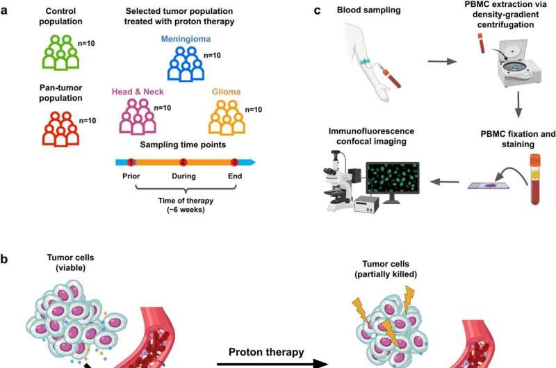
Overview of our platform to study the chromatin organization of peripheral blood mononuclear cells in a tumor and proton therapy setting. Credit: npj Precision Oncology (2023). DOI: 10.1038/s41698-023-00484-8
The ability to detect a developing tumor at a very early stage and to closely monitor the success or failure of cancer therapy is crucial for a patient’s survival. A breakthrough on both counts has now been achieved by researchers at the Paul Scherrer Institute PSI.
Researchers led by G.V. Shivashankar, head of PSI’s Laboratory for Nanoscale Biology and professor of Mechano-Genomics at ETH Zurich, were able to prove that changes in the organization of the cell nucleus of some blood cells can provide a reliable indication of a tumor in the body.
With their technique—using artificial intelligence—the scientists were able to distinguish between healthy and sick people with an accuracy of around 85 percent. Besides that, they managed to correctly determine the type of tumor disease—melanoma, glioma, or head and neck tumor. “This is the first time anyone worldwide has achieved this,” Shivashankar says. The researchers have published their results in the journal npj Precision Oncology.
Tumor cells give themselves away
It is normally very time-consuming to detect cancer in the body or to monitor it during the course of therapy, and these procedures are often carried out only in a late phase when the signs become obvious. For this reason, scientists engaged in fundamental research are looking for techniques that will be easy to use in everyday clinical practice as well as reliable and sensitive.
Enabling early detection of cancer

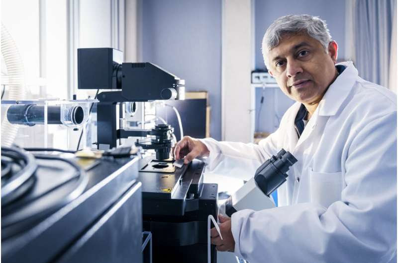
With his group’s new method and the use of artificial intelligence, G.V. Shivashankar hopes to improve tumour diagnosis. Credit: PAUL SCHERRER INSTITUTE/MARKUS FISCHER
Shivashankar’s research group set its sights on lymphocytes and monocytes, which people in the field refer to as mononuclear cells of peripheral blood. These are easy to obtain using a simple blood sample and have a round nucleus that is easy to see under the microscope. The researchers’ hunch was that the genetic material there reacts to substances released by the tumor into the bloodstream, the so-called secretome.
This activates the so-called chromatin in the nuclei of the blood cells and thus alters the organization of the genetic material it contains. This can then serve as either an indicator or a biomarker. “Our hypothesis was that the blood cells act as tumor detectors—and that took us a long way,” Shivashankar explains.
Artificial intelligence helps with diagnosis
The researchers used fluorescence microscopy to examine the chromatin of the blood cells—this is the term for the genetic material DNA packaged in a tangled ball. They recorded around 200 different characteristics in all, including the external texture, the packing density, and the contrast of the chromatin in the lymphocytes or monocytes.
They fed microscope images of samples from healthy and sick test participants into an artificial intelligence (AI) system. In doing so, they employed “supervised learning,” which is used to teach known differences in the software. In the subsequent “deep learning” approach, the algorithm itself identified differences between “healthy” and “sick” cells that would not be discernible to a human observer.
The research group took three different approaches. In an initial series of experiments, the researchers investigated whether the technique could distinguish healthy control subjects from those suffering from the disease. To do this, they compared the blood cells of ten cancer patients with those of ten healthy people. The artificial intelligence system was able to distinguish healthy and cancer patients with 85 percent accuracy.
“Even the analysis of just one arbitrary individual cell was carried out with a very high level of accuracy,” Shivashankar says. A second approach was to determine whether the AI system could distinguish between different types of tumors. To do this, the researchers fed the algorithm with the chromatin data from the blood cells of ten patients with a glioma (a tumor of the supporting tissue of nerve cells), a meningioma (a tumor of membranes that protect the brain and spinal cord), and a head and neck tumor.
This experiment, too, proved successful. The assignments had an accuracy of more than 85 percent. Finally, a third question concerned patients who were undergoing or had received treatment at the Centre for Proton Therapy CPT at PSI.
Damien Weber, director and Chief Physician of the ZPT, sees great potential in the diagnostic approach and asked 150 of his patients for permission to analyze their blood samples for the study, “We hope that the new method could improve both diagnosis and the monitoring of treatment success.”
To determine the success of the intervention, the scientists took blood samples before, during, and after the radiation therapy. Here, too, the software worked successfully and correctly assigned the patterns with a very high level of accuracy. The treatment was expected to reduce the concentration and build-up of tumor signals in the blood—this is what happened, and the appearance of the genetic material of the blood cells normalized.
“It was amazing to observe how the structure of the chromatin moved closer to the healthy pattern during the course of treatment,” says Shivashankar with satisfaction.
Many applications in tumor diagnosis and therapy are conceivable
From the point of view of the biologist and his colleagues, the new technique based on blood cell chromatin is applicable not only to the tumors they studied, but also to many different types of cancer. And it may not be limited to the follow-up of proton therapy, but rather applicable as well to many other forms of therapy, including radiation therapy in general, chemotherapy, and surgical operations.
Ascertaining whether this is true or not will require further research. As described in a paper in the journal Scientific Reports, Shivashankar’s group and collaborators at PSI’s Centre for Radiopharmaceutical Sciences (CRS) have already tested whether the chromatin biomarkers can be used to detect radiation- and chemo-resistant cells.
A lot of work remains to be done before regulatory authorities can approve the use of the new technique in clinical practice—particularly studies with a larger number of participants to ascertain how high the numbers of false positive alarms and false negatives are under clinical conditions. Shivashankar has no doubts about the path to clinical application or the prospect that patients will benefit from the technique, “The method stands,” he says.
More information: Kiran Challa et al, Imaging and AI based chromatin biomarkers for diagnosis and therapy evaluation from liquid biopsies, npj Precision Oncology (2023). DOI: 10.1038/s41698-023-00484-8
Journal information: npj Precision Oncology , Scientific Reports
Provided by Paul Scherrer Institute

Leave a Reply