by Matt Chittum, Virginia Tech

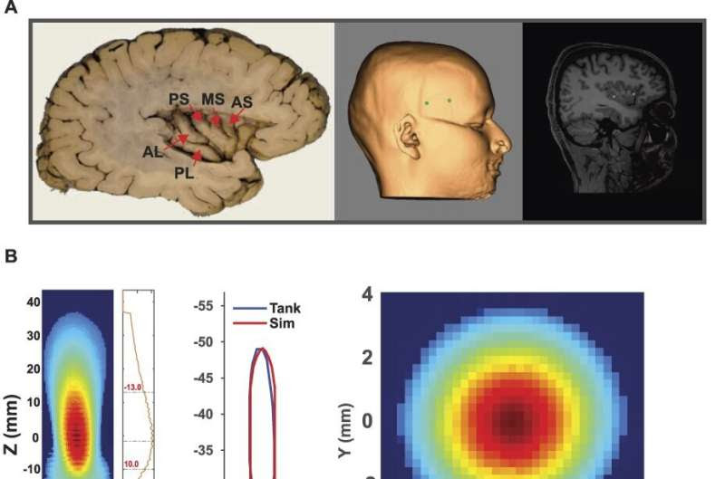
Targeting & acoustic modeling. Credit: Pain (2024). DOI: 10.1097/j.pain.0000000000003171
You feel a pain, so you pop a couple of ibuprofen or acetaminophen. If the pain is severe or chronic, you might be prescribed something stronger—an opioid pain killer that can be addictive under some circumstances.
But what if you could ease pain by non-invasively manipulating a spot inside your brain where pain is registered?
A new study by Wynn Legon, assistant professor at the Fralin Biomedical Research Institute at VTC, and his team points to that possibility. The study, published in the journal Pain, found soundwaves from low-intensity focused ultrasound aimed at a place deep in the brain called the insula can reduce both the perception of pain and other effects of pain, such as heart rate changes.
“This is a proof-of-principle study,” Legon said. “Can we get the focused ultrasound energy to that part of the brain, and does it do anything? Does it change the body’s reaction to a painful stimulus to reduce your perception of pain?”
Focused ultrasound uses the same technology used to view a baby in the womb, but it delivers a narrow band of sound waves to a tiny point. At high intensity, ultrasound can ablate tissue. At low-intensity, it can cause gentler, transient biological effects, such as altering nerve cell electrical activity
Neuroscientists have long studied how non-surgical techniques, such as transcranial magnetic stimulation, might be used to treat depression and other issues. Legon’s study, however, is the first to target the insula and show that focused ultrasound can reach deep into the brain to ease pain.
The study involved 23 healthy human participants. Heat was applied to the backs of their hands to induce pain. At the same time, they wore a device that delivered focused ultrasound waves to a spot in their brain guided by magnetic resonance imaging (MRI).
Participants rated their pain perception in each application on a scale of 0 to 9. Researchers also monitored each participant’s heart rate and heart rate variability—the irregularity of the time between heart beats—as a means to discern how ultrasound to the brain also affects the body’s reaction to a painful stimulus.
Participants reported an average reduction in pain of three-fourths of a point.
“That might seem like a small amount, but once you get to a full point, it verges on being clinically meaningful,” said Legon, also an assistant professor in the School of Neuroscience in Virginia Tech’s College of Science. “It could make a significant difference in quality of life, or being able to manage chronic pain with over-the-counter medicines instead of prescription opioids.”
The study also found the ultrasound application reduced physical responses to the stress of pain—heart rate and heart rate variability, which are associated with better overall health.
“Your heart is not a metronome. The time between your heart beats is irregular, and that’s a good thing,” Legon said. “Increasing the body’s ability to deal with and respond to pain may be an important means of reducing disease burden.”
The effect of focused ultrasound on those factors suggests a future direction for the Legon lab’s research—to explore the heart-brain axis, or how the heart and brain influence each other, and whether pain can be mitigated by reducing its cardiovascular stress effects.
More information: Wynn Legon et al, Noninvasive neuromodulation of subregions of the human insula differentially affect pain processing and heart-rate variability: a within-subjects pseudo-randomized trial, Pain (2024). DOI: 10.1097/j.pain.0000000000003171
Journal information: Pain
Provided by Virginia Tech

Leave a Reply