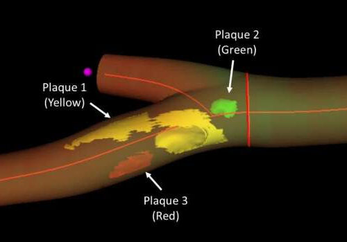
by Centro Nacional de Investigaciones Cardiovasculares Carlos III (F.S.P.) 3D model of a femoral artery obtained by real 3D imaging, revealing the location, number, and extent of distinct atherosclerotic plaques. In this sample, three plaques can be discerned (arrows), at the femoral bifurcation and distributed along the superficial branch. Credit: CNIC A new imaging technique...

