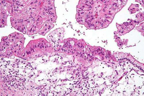
by H. Lee Moffitt Cancer Center & Research Institute Intermediate magnification micrograph of a low malignant potential (LMP) mucinous ovarian tumour. H&E stain. The micrograph shows: Simple mucinous epithelium (right) and mucinous epithelium that pseudo-stratifies (left – diagnostic of a LMP tumour). Epithelium in a frond-like architecture is seen at the top of image. Credit: Nephron /Wikipedia. CC...

