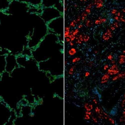
by Medical University of South Carolina Immunofluorescence images showing healthy (left) and scleroderma (right) lung. The scleroderma lung shows reduced levels of Cathepsin L (green) and increased levels of fibroblast activation marker (red). Image courtesy of Joe Mouawad, Medical University of South Carolina. Credit: Joe Mouawad, Medical University of South Carolina Much of the research...

