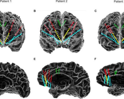
by Elsevier Intracranial recording electrodes sample depression-relevant prefrontal regions. (A-C) Frontal views of the reconstructed cortical surface and stereo-EEG recording contacts for Patient 1, Patient 2 and Patient 3, respectively. Stereo-EEG contacts are colored according to the gray matter region sampled: green, anterior cingulate cortex (ACC); red, dorsolateral prefrontal cortex (dlPFC); blue, orbitofrontal cortex (OFC);...

