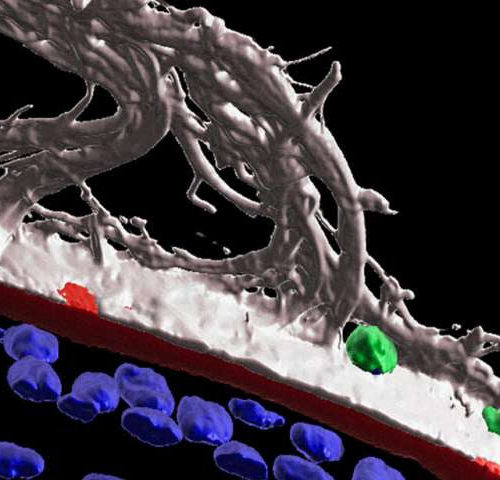
by Thomas Jefferson University 3D surface structure imaging at one day post-corneal wounding shows immune cells (CD45+, green) migrating along within ciliary zonule fibrils (MAGP1+, white) that extend along the surface of the matrix capsule that surrounds the lens (perlecan+, red). Also seen are the ciliary zonules (white) that link the lens to the ciliary...
Tag: <span>cornea cells</span>
Post
Study shows stem cells constitute alternative approach for treating corneal scarring
Durham, NC – Infection, inflammation, trauma, disease, contact lenses – all of these and more can lead to corneal scarring, which according to the World Health Organization is a leading cause of blindness worldwide. While corneal transplant remains the gold standard to treat this condition, patient demand far outweighs donor supply. However, in a study...
Post
Stanford lab grows cornea cells for transplant
PALO ALTO — A Stanford research team has created a potentially powerful new way to fix damaged corneas — a major source of vision problems and blindness. Millions of new eye cells are being grown in a Palo Alto lab, enlisting one of medicine’s most important and promising new tools: refurbishing diseased and damaged tissue...


