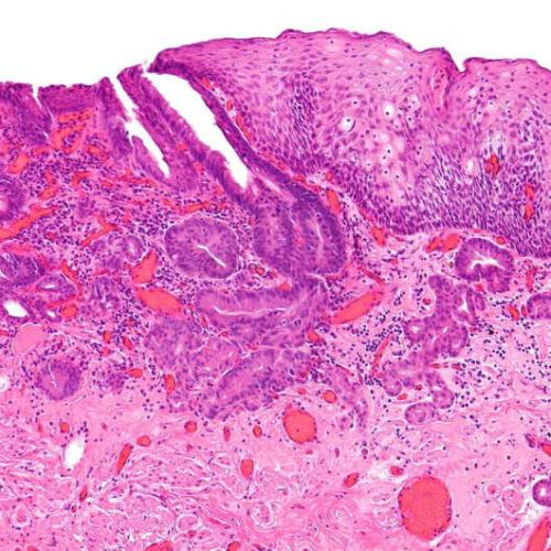
by Caslon Hatch, Johns Hopkins University Intermediate magnification micrograph of an intramucosal esophageal adenocarcinoma, a type of esophageal cancer. H&E stain. Endoscopic mucosal resection specimen. The images show normal squamous epithelium (right of image) and an adenocarcinoma (left of image). The adenocarcinoma has a typical morphology; it is composed of cohesive clusters of cells arranged...

