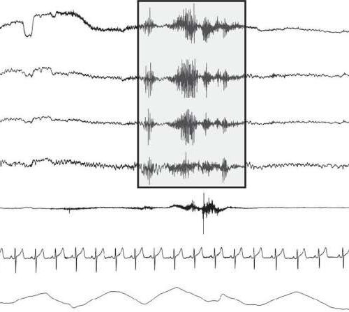
by American Physiological Society Figure 1. A representative image showing a 20-s polysomnography (PSG) sleep tracing including electrooculography (EOG), electroencephalography (EEG; F3, C3, and O1 leads), chin electromyography (EMG), electrocardiogram (ECG), and respiration (Resp). The light gray box outlines an exemplary cortical arousal as marked by an abrupt shift in the EEG frequency. Credit: DOI:...

