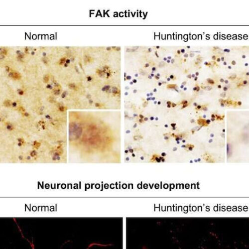
by National Research Council of Science & Technology Differences in FAK activation and neuronal protrusion formation in brain tissues of normal and Huntington’s disease patients. Credit: Korea Institute of Science and Technology (KIST) Huntington’s disease (HD) is a hereditary brain disease caused by a mutation in the huntingtin gene. HD is a neurodegenerative disease without...
Tag: <span>Huntington’s Disease</span>
Establishing a novel strategy to tackle Huntington’s disease
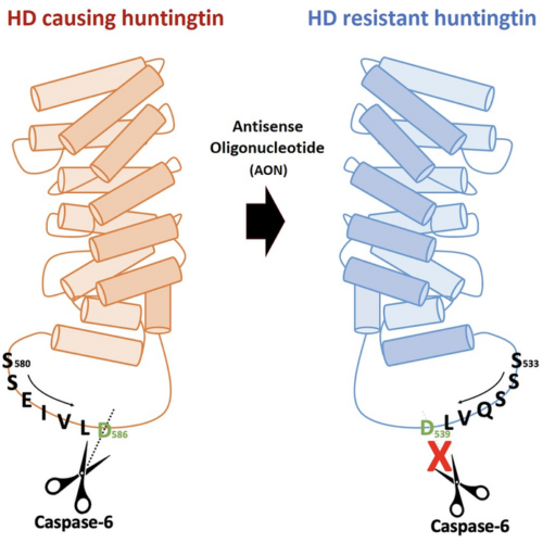
THE KOREA ADVANCED INSTITUTE OF SCIENCE AND TECHNOLOGY (KAIST) IMAGE: FIGURE. HUNTINGTON’S DISEASE RESISTANCE HUNTINGTIN PROTEIN INDUCED BY ANTISENSE OLIGONUCLEOTIDE (AON) IS RESISTANT TO CASPASE-6 CLEAVAGE, THEREFORE, DOES NOT CAUSE HUNTINGTON’S DISEASE WHILE MAINTAINING NORMAL FUNCTIONS OF HUNTINGTIN. CREDIT: HYEOUNGJUN KIM, ET AL., 2022 Through an international joint research effort involving ProQR Therapeutics of the...
New system that defines Huntington’s disease will ‘revolutionize’ drug trials
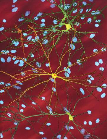
by University College London A montage of three images of single striatal neurons transfected with a disease-associated version of huntingtin, the protein that causes Huntington’s disease. Nuclei of untransfected neurons are seen in the background (blue). The neuron in the center (yellow) contains an abnormal intracellular accumulation of huntingtin called an inclusion body (orange). Credit:...
Medication benefits patients with Huntington’s disease-associated chorea
UNIVERSITY OF TEXAS HEALTH SCIENCE CENTER AT HOUSTON IMAGE: ERIN FURR STIMMING, MD, PROFESSOR OF NEUROLOGY WITH MCGOVERN MEDICAL SCHOOL AT UTHEALTH HOUSTON AND DIRECTOR OF THE HUNTINGTON’S DISEASE SOCIETY OF AMERICA CENTER OF EXCELLENCE AT UTHEALTH HOUSTON NEUROSCIENCES. CREDIT: PHOTO BY UTHEALTH HOUSTON A new drug has proven beneficial in treating a movement disorder...
Huntington’s disease: Astrocytes to the rescue !
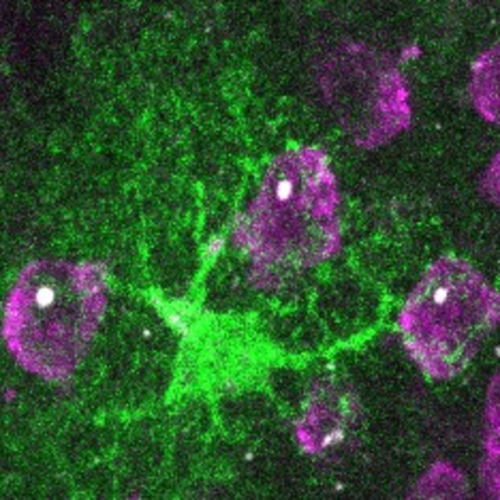
CNRS IMAGE: AN ASTROCYTE (GREEN) IS SURROUNDED BY NEURONS (MAGENTA) EACH CONTAINING AN AGGREGATE OF MUTATED HUNTINGTIN (WHITE). CREDIT: © LAURENE ABJEAN Huntington’s disease1 is caused by a mutation in the Huntingtin gene, a protein necessary for the proper functioning of several brain cells. Mutated, it is no longer able to perform properly: it can even...
Team develops new mouse model to shed light on the mystery surrounding Huntington’s disease onset
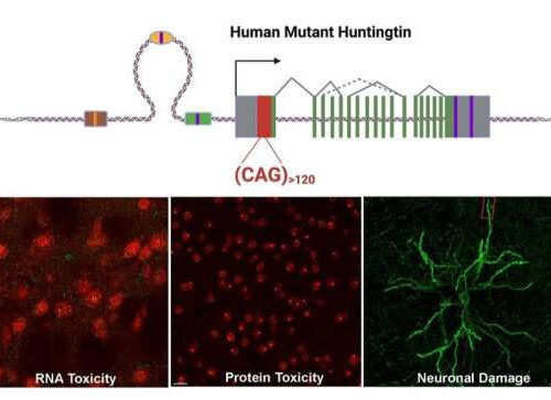
by University of California, Los Angeles The upper portion of the schematic illustrates generation of a novel mouse model of Huntington’s disease with long, uninterrupted CAG repeats. In the new model, the striatum shows RNA and protein toxicities originated from the pure CAG repeats, which lead to neuronal damage. Credit: UCLA Health/Yang Lab Researchers at...
Retrotransposon expression is repressed in Huntington’s disease
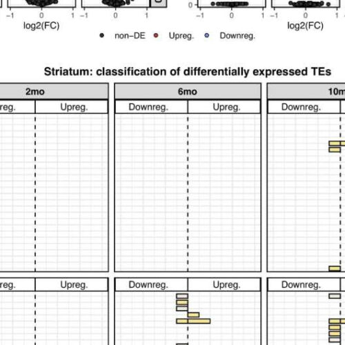
by John Hewitt, Medical Xpress Figure 1. Differentially expressed TEs. (A) Volcano plot showing the murine TE consensus sequences differentially expressed in the striatum of 2-, 6-, and 10-mo mice in Q111 and Q175 genotypes compared to control (Q20). Blue color indicates the TE consensus sequences that result downregulated in each tested comparison (Q111 vs....
Gene Therapy for the Treatment of Huntington’s Disease
MARY ANN LIEBERT, INC./GENETIC ENGINEERING NEWS IMAGE: THE FIRST PEER-REVIEWED JOURNAL IN THE FIELD AND PROVIDES ALL-INCLUSIVE ACCESS TO THE CRITICAL PILLARS OF HUMAN GENE THERAPY: RESEARCH, METHODS, AND CLINICAL APPLICATIONS. CREDIT: MARY ANN LIEBERT INC., PUBLISHERS Gene therapy targeting the messenger RNA (mRNA) of the mutated huntingtin gene (HTT) can provide long-lasting therapeutic benefit...
CRISPR-Cas13 technique targets proteins causing ALS and Huntington’s disease in the mouse nervous system
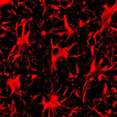
by Liz Ahlberg Touchstone, University of Illinois at Urbana-Champaign Spinal cord astrocytes, the cells seen in this fluorescent microscope image, are involved in the progression of ALS. A new CRISPR-Cas13 system targeting mutant protein production in these cells improved outcomes for mice with ALS. Credit: Thomas Gaj and Colin Lim A single genetic mutation can...
Huntington’s disease: The ultrastructure of huntingtin inclusions revealed
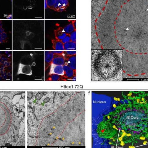
by Ecole Polytechnique Federale de Lausanne Fig. 1: Confocal microscopy and CLEM revealed the ring-like structure of the Httex1 72Q inclusions in HEK cells. a Epitope mapping of the Httex1 antibodies used in this study. b Confocal imaging of Httex1 72Q inclusions formed 48 h after transfection in HEK cells. All the Htt antibodies showed strong immunoreactivity to the...

