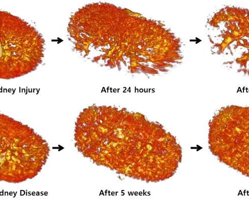
by Pohang University of Science and Technology Vascular changes in acute and diabetic renal failure. Credit: POSTECHA research team at POSTECH (Pohang University of Science and Technology) has investigated kidney diseases using ultrafast ultrasound that captures 1,000 images in just one second. The research team has achieved imaging of the three-dimensional microvasculature of the kidneys using...
Tag: <span>Imaging</span>
Lighting the way to selective membrane imaging
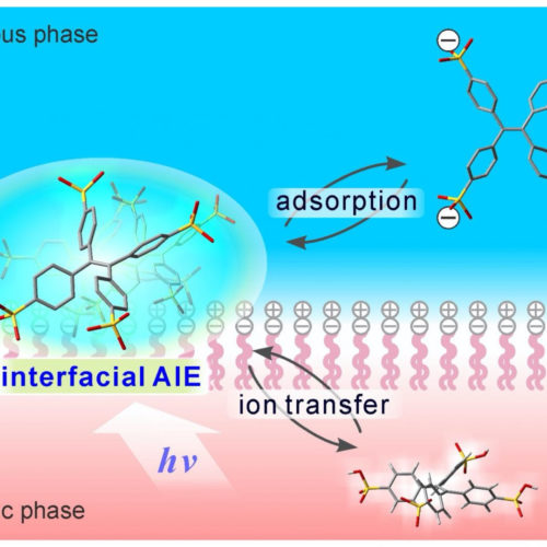
KANAZAWA UNIVERSITY IMAGE: WATER-SOLUBLE TETRAPHENYLETHENE (TPE) DERIVATIVES BEARING ANIONIC GROUPS EXHIBIT AGGREGATION-INDUCED EMISSION (AIE) BEHAVIOR SPECIFICALLY AT LIQUID-LIQUID INTERFACES. INTERFACIAL AIE PROCESS RESPONDS REVERSIBLY TO THE EXTERNALLY APPLIED POTENTIAL AT A BIOMEMBRANE-MIMETIC INTERFACE. Kanazawa, Japan – Researchers at Kanazawa University monitored the emission of blue-green light from water-soluble tetraphenylethene molecules adsorbed at a phospholipid-adsorbed liquid-liquid interface made...
Imaging agent developed at Washington University spotlights inflammation
WASHINGTON UNIVERSITY SCHOOL OF MEDICINE Many of the most common diseases — cancer, diabetes, cardiovascular and lung disease, and even COVID-19 — have been linked to chronic or excessive inflammation. Blood tests can indicate that some part of a person’s body is inflamed, but doctors don’t have a good way to zero in on the...
Brain imaging expertise supports new discoveries on decision-making process
by Toby Leigh, University of Plymouth Research carried out by a University academic has shed new light on the fundamentals of how, and why, we make the decisions we do. In two separate studies, UKRI Future Leader Fellow and Lecturer in Psychology, Dr. Elsa Fouragnan has used her expertise in functional magnetic resonance imaging (fMRI)...
Study finds evidence for existence of elusive ‘metabolon’
UNIVERSITY PARK, Pa. — For more than 40 years, scientists have hypothesized the existence of enzyme clusters, or “metabolons,” in facilitating various processes within cells. Using a novel imaging technology combined with mass spectrometry, researchers at Penn State, for the first time, have directly observed functional metabolons involved in generating purines, the most abundant cellular...
IMAGING TECHNIQUE SPOTS COLORECTAL TUMORS WITH 100% ACCURACY
BETH MILLER-WUSTL A new imaging technique in development provides accurate, real-time, computer-aided diagnosis of colorectal cancer, researchers say. Using deep learning, a type of machine learning, researchers used the technique on more than 26,000 individual frames of imaging data from colorectal tissue samples to determine the method’s accuracy. Compared with pathology reports, the method identified...
‘Mindful people’ feel less pain; MRI imaging pinpoints supporting brain activity
WINSTON-SALEM, NC – Sept. 6, 2018 – Ever wonder why some people seem to feel less pain than others? A study conducted at Wake Forest School of Medicine may have found one of the answers – mindfulness. “Mindfulness is related to being aware of the present moment without too much emotional reaction or judgment,” said...
- 1
- 2


