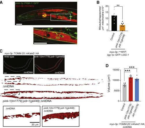
by Erik de Wit, University of Queensland (A) Representative fluorescent image of the PINK-1::GFP translational reporter in a myo-3p::mCherry background. The white box is magnified in the panel below, and body wall muscle cells are indicated by yellow dashed lines. Scale bar, 10 μm. (B) Quantification of mitophagy in body wall muscle cells. Columns are means...

