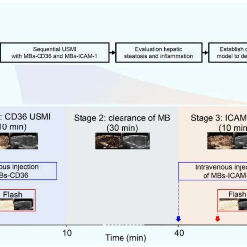
by KeAi Communications Co. Animal study flowchart and schematic illustration of sequential USMI. Credit: Liver Research (2023). DOI: 10.1016/j.livres.2023.11.002Non-alcoholic fatty liver disease (NAFLD), recently renamed metabolic dysfunction-associated fatty liver disease (MAFLD), is spectrum of diseases that ranges from non-alcoholic fatty liver (NAFL) to non-alcoholic steatohepatitis (NASH). Invasive liver biopsy continues to be the gold standard...
Tag: <span>ultrasound imaging</span>
A New Vision for Ultrasound Imaging
Ultrasound research specialist and 2023 MIT Excellence Award winner Nicole Henning adapts ultrasound technology for more sensitive, less invasive imaging for disease modeling. Nicole Henning did not foresee becoming an expert in ultrasound imaging. Before joining Koch Institute for Integrative Cancer Research at MIT as an ultrasound research specialist at the Preclinical Imaging and Testing (P&IT) Core...
AI-powered ultrasound imaging that detects breast cancer
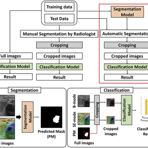
POHANG UNIVERSITY OF SCIENCE & TECHNOLOGY (POSTECH) IMAGE: RESEARCH IMAGE CREDIT: POSTECH Breast cancer undisputedly has the highest incidence rate in female patients. Moreover, out of the six major cancers, it is the only one that has shown an increasing trend over the past 20 years. The chance of survival would be higher if breast...
Ultrasound imaging can help solve the Alzheimer’s disease “chicken and egg” problem
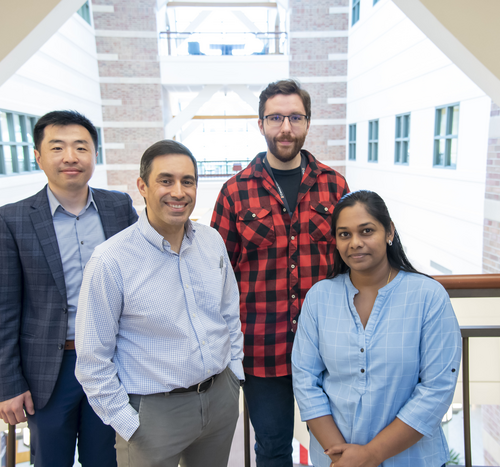
BECKMAN INSTITUTE FOR ADVANCED SCIENCE AND TECHNOLOGY IMAGE: (FROM LEFT) PENGFEI SONG, DR. DAN LLANO, MATTHEW LOWERISON, AND NATHIYA CHANDRA SEKARAN ARE PART OF THE BECKMAN INSTITUTE FOR ADVANCED SCIENCE AND TECHNOLOGY RESEARCH TEAM TO RECEIVE FEDERAL FUNDING TO DEVELOP ULTRASOUND IMAGING METHODS FOR STUDYING THE NEUROVASCULAR CHANGES UNDERLYING ALZHEIMER’S DISEASE. CREDIT: BECKMAN INSTITUTE FOR...
Major advance in 3D ultrasound imaging to observe entire organs
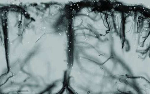
by ESPCI Paris Credit: Alexandre Dizeux Two successive studies by the Physics for Medicine Paris laboratory (ESPCI Paris-PSL, Inserm, CNRS) highlight advances in non-invasive 3D ultrasound imaging, making it possible to observe blood flow in real time in two whole organs, the heart and the brain. This work was published in JACC Cardiovascular Imaging and featured on the...
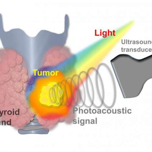
Thyroid cancer now diagnosed with AI photoacoustic/ultrasound imaging
by Pohang University of Science & Technology A schematic diagram of acquiring the photoacoustic signal generated when laser light is irradiated to a malignant thyroid nodule with an ultrasonic sensor. Credit: POSTECH A lump in the thyroid gland is called a thyroid nodule, and 5-10% of all thyroid nodules are diagnosed as thyroid cancer. Thyroid...

