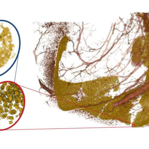
by Göttingen University The image shows neuronal cell nuclei of the dentus gyratus (yellow) and associated blood vessels (red). By varying the magnification of the X-ray optics, one can “zoom in” on the densely packed band of neurons (in the red oval) and also resolve the substructure of the cell nucleus (blue oval). The study...

