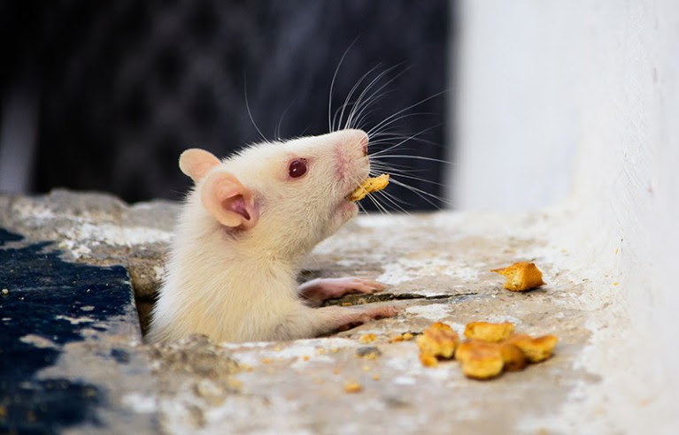
Scientists found the cells in mice — and say they could lead to a better understanding of human appetite.
Carissa Wong

Credit: Nishant Sharma/Getty
Brain cells that control how quickly mice eat, and when they stop, have been identified. The findings, published in Nature1, could lead to a better understanding of human appetite, the researchers say.
Nerves in the gut, called vagal nerves, had already been shown to sense how much mice have eaten and what nutrients they have consumed2. The vagal nerves use electrical signals to pass this information to a small region in the brainstem that is thought to influence when mice, and humans, stop eating. This region, called the caudal nucleus of the solitary tract, contains prolactin-releasing hormone neurons (PRLH) and GCG neurons. But, until now, studies have involved filling the guts of anaesthetized mice with liquid food, making it unclear how these neurons regulate appetite when mice are awake.
To answer this question, physiologist Zachary Knight at the University of California, San Francisco, and his colleagues implanted a light sensor in the brains of mice that had been genetically modified so that the PRLH neurons released a fluorescent signal when activated by electrical signals transmitted along neurons from elsewhere in the body. Knight and his team infused a liquid food called Ensure — which contains a mixture of fat, protein, sugar, vitamins and minerals — into the guts of these mice. Over a ten-minute period, the neurons became increasingly activated as more of the food was infused. This activity peaked a few minutes after the infusion ended. By contrast, the PRLH neurons did not activate when the team infused saline solution into the mice’s guts.
When the team allowed the mice to freely eat liquid food, the PRLH neurons activated within seconds of the animals starting to lick the food, but deactivated when they stopped licking. This showed that PRLH neurons respond differently, depending on whether signals are coming from the mouth or the gut, and suggests that signals from the mouth override those from the gut, says Knight.
By using a laser to activate PRLH neurons in mice that were eating freely, the researchers could reduce how quickly the mice ate. Further experiments showed that PRLH neurons did not activate during feeding in mice that lacked most of their ability to taste sweetness, suggesting that taste activated the neurons.
The researchers also found that GCG neurons are activated by signals from the gut, and control when mice stop eating. “The signals from the mouth are controlling how fast you eat, and the signals from the gut are controlling how much you eat,” says Knight.
“I’m extremely impressed by this paper,” says neuroscientist Chen Ran at Harvard University in Boston, Massachusetts. The work provides original insights on how taste regulates appetite, he says. The findings probably apply to humans, too, Ran adds, because these neural circuits tend to be well conserved across both species.
doi: https://doi.org/10.1038/d41586-023-03679-y
References
Ly, T. et al. Nature https://doi.org/10.1038/s41586-023-06758-2 (2023).
Leave a Reply