by Emily Caldwell, The Ohio State University

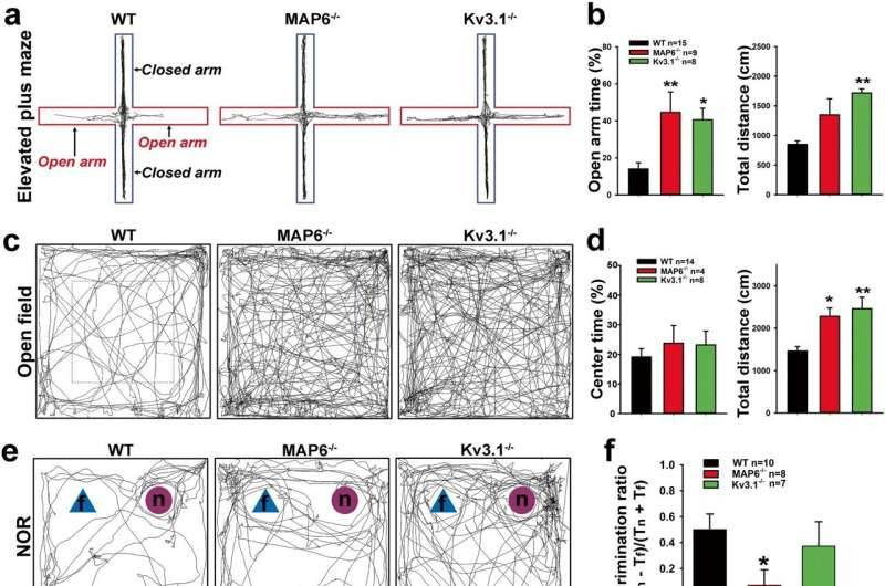
Behavioral alterations in MAP6−/− and Kv3.1−/− mice. Adult (3–6 months old) WT B6 (black bars), MAP6−/− (red bars), and Kv3.1−/− (green bars) mice were used in a series of behavioral assays. Each group contained approximately half male and half female mice. a Example traces in the elevated plus maze (EPM) with open arms indicated in red and closed arms in dark blue. b Summaries of open arm time (100 × TOpen/(TOpen + TClosed); WT control, MAP6−/− p = 0.001, Kv3.1−/− p = 0.029) (left) and total travel distance (cm; WT control, MAP6−/− p = 0.088, Kv3.1−/− p = 0.003)(right) in EPM. c Example traces in the open field test with blue dashed lines to indicate the center. d Summaries of the center time percentage (100 × TCenter/TTotal; Overall p = 0.595)(left) and the total travel distance (cm; WT control, MAP6−/− p = 0.045, Kv3.1−/− p = 0.002)(right) in the open field test. e Example traces in the novel object recognition (NOR) test with the blue triangles as the familiar objects (f) and the red circles as the novel objects (n). f Summary of the discrimination ratio, (Tn – Tf)/(Tn + Tf). WT control, MAP6−/− p = 0.044, Kv3.1−/− p = 0.096. g Summary of the rotarod test. In trial #3, MAP6−/− p = 0.041. In trial #4, MAP6−/− p = 0.001. h Summary of the balance beam test. i Summary of spontaneous alternation in the Y maze test (overall p = 0.801). Each result is provided as mean ± SEM. Mouse numbers are provided in the figure. One-Way ANOVA followed by Dunnett’s test: *, p < 0.05; **, p < 0.01. Credit: Molecular Psychiatry (2023). DOI: 10.1038/s41380-023-02286-7
The discovery of a physical interaction between two proteins in brain cells that can be traced in mice to control of movement, anxiety and memory could one day open the door to development of new schizophrenia treatment strategies, researchers say.
The research group is the first to determine that the two proteins, both among the dozens of proteins related to risk for the development of schizophrenia, bind to each other under normal conditions in multiple regions of the brain, and that their connection was found in mice to be key to maintaining normal movement, memory function and anxiety regulation.
When that connection doesn’t happen as it should, they found, behavior can be negatively affected—in mice, disruption to the proteins’ ability to interact increased hyperactivity, reduced risk avoidance and impaired memory. Though delusions and hallucinations are hallmark symptoms of schizophrenia, the condition also encompasses additional symptoms, including movement and memory problems.
“These two proteins are seemingly unrelated, and our study has provided a link between them that wasn’t recognized before,” said lead author Chen Gu, associate professor of biological chemistry and pharmacology in The Ohio State University College of Medicine.
“There are more than 100 genes that have been identified as risk genes for schizophrenia, but we still don’t know the real mechanisms behind those risks,” Gu said. “We’re hopeful that getting a better understanding of this mechanism could help in the long run to find a new treatment that could benefit patients with schizophrenia.”
The study was published recently in the journal Molecular Psychiatry.
Previous post-mortem studies have identified risk genes for schizophrenia based on signs of protein dysfunction detected in brain tissue. Among them are the proteins in this study: MAP6, which has a role in supporting a neuron’s cytoskeleton or, more specifically, microtubules, and Kv3.1, which helps control the maximal frequency of electrical signaling by neurons.
Gu’s lab has studied Kv3.1 for many years, often working with genetically altered mice lacking its gene. As the team began exploring a connection between Kv3.1 and MAP6, first study author Di Ma, a graduate student in the lab, found that mice lacking the genes for both proteins experienced similar behavior changes.
“That’s how we started looking at their relationship in more detail,” Gu said.
In this study, Ma and her lab mates took a more nuanced look at how the proteins’ connection relates to behavior by disrupting their ability to bind to each other in specific brain regions in mice: the hippocampus, which governs learning and memory, and the nearby amygdala, where emotions are processed.
The researchers found that disruption to the proteins’ connection in the amygdala led to a reduction in risk avoidance—shown in mice as a lack of fear of height. Blocking the proteins’ attachment in the hippocampus resulted in hyperactivity and lower recognition of a familiar object. Though some behavior changes in these experiments differed from the longer list of changes seen in mice completely lacking one or both genes, the finding provided important insights about where the proteins’ interactions, or lack thereof, have the strongest effect on behavior.
“Different physiological functions we are engaged in daily are governed by different brain regions,” Gu said. “That’s an advance provided by our study—because previously we only knew global knockout mice had these behavioral alterations, we didn’t really know what brain region was responsible for them.”
The next step in Gu’s lab will be exploring any links between social behavior in mice and these proteins’ functions in the prefrontal cortex, a brain region important to decision making and planning.
In a series of biochemistry and cell biology experiments, the researchers also determined how the proteins bind and how that connection affects their positioning inside neurons. Results showed MAP6 stabilizes the Kv3.1 channel in a specific type of interneurons, where it helps these cells keep brain signals at an even keel. A drop in the expression of MAP6, on the other hand, dramatically decreased the level of Kv3.1 in those interneurons.
The combined findings suggest that when the proteins don’t bind properly, there isn’t enough Kv3.1 available to maintain interneurons’ signal-control function, leading to an imbalance of neural inhibition and excitation in affected brain regions—and related negative behavioral symptoms. This type of interneurons, capable of generating nerve impulses in high frequencies, represent a key therapeutic target for schizophrenia.
“Our study further provides a link between the MAP6 dysfunction and the interneuron signal dysfunction, and we now know that there are two proteins that interact and that one could alter the other,” Gu said. “That opens up potential new directions for treatment strategies.”
More information: Di Ma et al, A cytoskeleton-membrane interaction conserved in fast-spiking neurons controls movement, emotion, and memory, Molecular Psychiatry (2023). DOI: 10.1038/s41380-023-02286-7
Journal information: Molecular Psychiatry
Provided by The Ohio State University

Leave a Reply