September 3, 2024
by Ingrid Fadelli , Medical Xpress

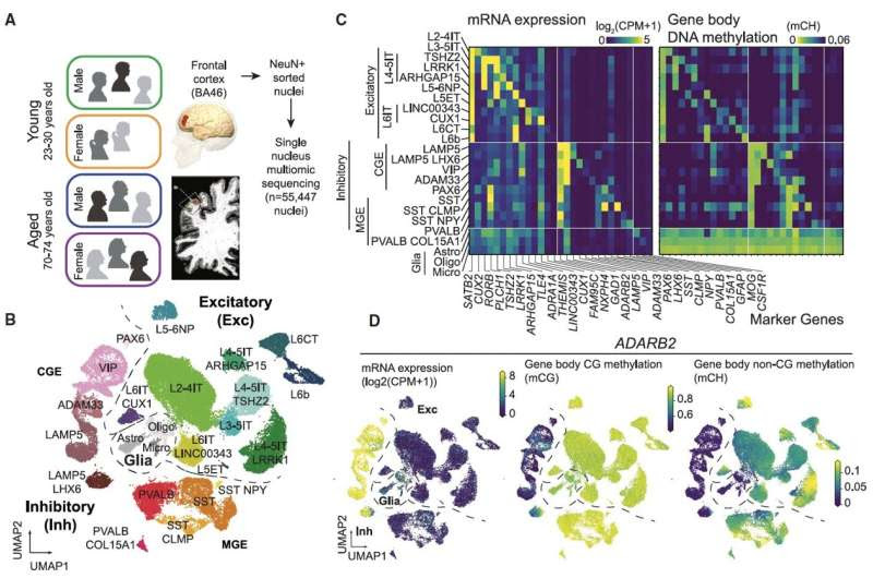
(A) Study design. (B) UMAP embedding of nuclei assigned to 11 excitatory, 10 inhibitory, and 3 glial types using DNA methylation in 100 kb bins. (C) RNA expression and gene body DNA methylation at non-CG sites (mCH) for cell type marker genes. (D) Correspondence of mRNA, mCG, and mCH for the CGE interneuron marker gene, ADARB2. Credit: Neuron (2024). DOI: 10.1016/j.neuron.2024.05.013
Aging is known to have profound effects on the human brain, prompting changes in the composition of cells and the expression of genes, while also altering aspects of the interaction between genes and environmental factors. While past neuroscience studies have pinpointed many of the molecular changes associated with aging, the age-related genetic factors influencing specific neuron populations remains poorly understood.
Recent studies on flies, mice, primates and human brain tissue utilizing single-cell or single-nucleus RNA-sequencing and genetic experimental techniques shed new light on these cell-type-specific changes. For instance, they unveiled the effects of aging on glial cells in the mouse and human brain, associations between cell-specific changes and modified chromatin proteins, and the influence of DNA methylation in the aging of various tissues.
Researchers at University of California (UC) San Diego and Salk Institute recently carried out a study aimed at better understanding how both age and sex impact human cortical neurons at a single-cell level. Their findings, published in Neuron, offer new insights into how aging affects cell composition, gene expression and DNA methylation across human brain cell types, while also uncovering differences between gene expression and DNA methylation in females and males.
“Altered transcriptional and epigenetic regulation of brain cell types may contribute to cognitive changes with advanced age,” Jo-Fan Chien, Hanqing Liu and their colleagues wrote in their paper. “Using single-nucleus multi-omic DNA methylation and transcriptome sequencing (snmCT-seq) in the frontal cortex from young adult and aged donors, we found widespread age- and sex-related variation in specific neuron types.”
The researchers examined prefrontal cortex neurons in post-mortem tissue from the brain of young adults (aged 23 to 30 years old) and older adults (aged 70 to 74 years old) of both sexes. They analyzed these neurons using a genetic technique known as single-nucleus multi-omic DNA methylome and transcriptome sequencing (snmCT-seq).
The team compiled a large dataset containing profiles for more than 55,000 cells extracted from a total of 11 human donors. While some cell processes were found to be stable across all individuals, others appeared to vary depending on a person’s age and sex.
“The proportion of inhibitory SST (somatostin)- and VIP (vasoactive intestinal polypeptide)-expressing neurons was reduced in aged donors,” wrote Chien, Liu and their colleagues. “Excitatory neurons had more profound age-related changes in their gene expression and DNA methylation than inhibitory cells.
“Hundreds of genes involved in synaptic activity, including EGR1, were less expressed in aged adults. Genes located in subtelomeric regions increased their expression with age and correlated with reduced telomere length.”
The analyses carried out by the researchers revealed that the expression of genes implicated in synaptic function (i.e., supporting communication between neurons via synapses) decreased with age. The downregulation of these genes was found to be accompanied by an increase in DNA methylation, an epigenetic modification that entails the addition of a methyl group to human DNA.
The researchers also observed notable sex differences in both the expression of genes and DNA methylation. While the frontal cortex of male and female donors contained the same types of cells, they found that some genes were more expressed in males (e.g., PDIA2) and genes that were more expressed in females (e.g., RNA LINC01115).
Typically, females undergo a process known as X inactivation, which entails the silencing of one of the X chromosomes to align gene expression with that of males (who only have one X chromosome). Yet some genes may escape this silencing and be expressed by both of a female’s X chromosomes.
The researchers were able to identify DNA methylation patterns pointing to the X-chromosome-associated genes that escape X inactivation. One of these genes is GEMIN8, which was found to be active in some cell types despite the overall inactivation of one X chromosome.
“We mapped cell-type-specific sex differences in gene expression and X-inactivation escape genes,” wrote Chien, Liu and their colleagues.
Overall, this study sheds further light on the cell-type specific effects of age and sex on human cortical neurons. In the future, the experimental methods employed by these researchers could be used to analyze brain tissues extracted from a wider pool of female and male donors of different ages, as this could help to validate their findings and potentially lead to new interesting discoveries.
More information: Jo-Fan Chien et al, Cell-type-specific effects of age and sex on human cortical neurons, Neuron (2024). DOI: 10.1016/j.neuron.2024.05.013
Journal information: Neuron
© 2024 Science X Network

Leave a Reply